English
 Deutsch
  Nederlands
3-D live MRI-Prostate biopsy – the optimized further development to the classical biopsy
When is a biopsy necessary?
A biopsy is necessary whenever there is a sound reason to suspect prostate cancer.
- 3-D live MRI-Prostate biopsy – the optimized further development to the classical biopsy
- When is a biopsy necessary?
- Frontal Plane of the Prostate
- Maximum Assurance with the Alta Klinik Biopsy
- The Alta Klinik MRI-Guided Biopsy Procedure
- The prostate biopsy carried out at the Alta Klinik is painless.
- Access Route to the Prostate with the Alta Klinik Biopsy
- A Sedative Instead of General Anesthesia
- A Sterile and Hygienic Procedure
- Uncomplicated Prostate Biopsy Procedure
- Prerequisite for a Targeted Prostate Biopsy
The only way to clarify a case of suspected prostate cancer is to have a pathologist examine the relevant tissue samples. This applies also to the degree of malignancy (Gleason score) that a given tumor might have. The tissue samples are removed in a procedure that is referred to as a biopsy. There is no other way to know for sure.
You are currently viewing a placeholder content from YouTube. To access the actual content, click the button below. Please note that doing so will share data with third-party providers.
More InformationFrontal Plane of the Prostate
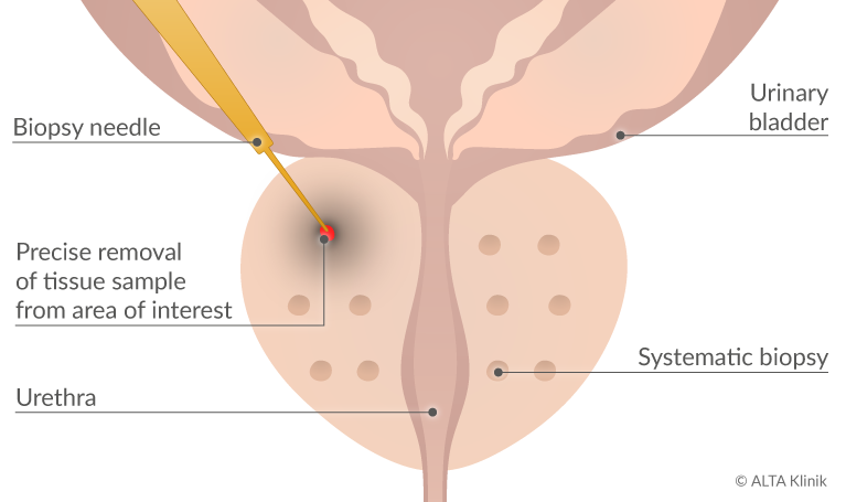
- Targeted removal of tissue samples from a suspicious site
- Systematic removal of tissue samples from the prostate
Maximum Assurance with the Alta Klinik Biopsy
With our MRI-guided prostate biopsy we can take tissue samples from suspicious sites from all areas of the prostate. One reason for this is that we use a sterile way in the gluteal region (transgluteal) and therefore have no restrictions on the accessibility of suspicious sites. On the other hand, because our biopsy is MRI-guided and takes place via live images in the MRI-scanner, which can depict a suspicious site. For safety reasons and in accordance with the S3 guidelines for Prostate Cancer, we take targeted samples from suspicious sites as well as systematic tissue samples from the prostate. The combination of precisely targeted and systematic tissue samples offers our patients a maximum degree of assurance.
Advantages of the ALTA Klinik Biopsy
- the combination biopsy of the ALTA Clinic provides the patient with maximum safety because suspicious sites can be biopsies and systematic samples can also be taken from the prostate
- the biopsy is performed using current live images under MRI control and not only based on MRI images taken days or weeks previously
- suspicious areas can be biopsied with a targeted hit rate of 99%
- the biopsy is painless
- access to the prostate is not via the colon or perineum, but above the buttock region (transgluteal)
- no preventive antibiotic is taken because the biopsy runs over the upper buttock region and therefore there is no risk of infection from intestinal bacteria
- according to urological recommendations (S 3 guideline for prostate carcinoma), systematic tissue samples are additionally taken from the prostate gland
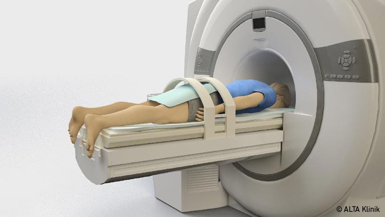
- for the biopsy, the patient does not sit with his legs apart on a urological chair, but on the MRI table lying on his stomach with his legs closed.
- no contact to the genital area
- no anaesthesia required
- no inpatient stay necessary, outpatient only
- the patient has no pain or physical limitations after the biopsy
- the biopsy is performed safely and quickly because a professional and experienced team at the ALTA Klinik has standardized the procedures over many years
The Alta Klinik MRI-Guided Biopsy Procedure
The procedure is carried out after the exact location of suspicious tissue has already been determined on the basis of an MRI scan of the prostate.
In the context of the biopsy itself, we first guide the needle with the help of the images generated by the MRI device to the exact location of the suspicious site or sites in the prostate. We then proceed to remove a tissue sample from the targeted site only after we have precisely lined up both the point of the needle and the suspicious tissue in the real-time images.
After the targeted removal of tissue samples from the identified sites, we remove additional tissue samples according to a systematic method that is specified in the latest guidelines for physicians in Germany.
The prostate biopsy carried out at the Alta Klinik is painless.
Thanks to the prior administration of a local anesthetic at the access site and at the prostate capsule, the biopsy procedure is painless for the patient. Moreover, our patients usually remain free of pain and physical limitations after their biopsies.
Access Route to the Prostate with the Alta Klinik Biopsy
Conventional biopsy procedures involve accessing the prostate either through the rectum or the perineum. Instead, we use an approach that begins relatively far away from the rectum, namely, at a location in the upper buttocks marking the transition from the lower back to the buttocks. In contrast to a transrectal biopsy, the point of access we use makes it unnecessary to administer an antibiotic because there is no risk of infection from intestinal bacteria.
Moreover, our access route enables us to reach all locations within the prostate gland, including locations that offer little room for maneuvering, or that are positioned on the periphery, at the front, at the rear, at the top or at the bottom of the prostate. These locations are often very difficult or impossible to reach when using the rectum as a point of entry.
A Sedative Instead of General Anesthesia
Our patients are given a sedative to make sure they are able to remain still while in a prone position. Otherwise, patient movements would likely make it necessary to repeatedly correct the progress of the needle on its way to the sites of suspicious tissue within the prostate. The administration of a sedative comes with a number of standard precautions such as refraining from operating a motor vehicle. We inform our patients both orally and in writing about the precautions that are necessary after taking a sedative.
A Sterile and Hygienic Procedure
We perform our biopsies under sterile and hygienic conditions. The entry site is covered with a sterile bandage and only disposable articles are used.
Uncomplicated Prostate Biopsy Procedure
Our patients benefit from a fast and uncomplicated biopsy procedure that is carried out in an outpatient setting and therefore does not require admission to a hospital.
Moreover, our patients face no direct physical restrictions after the procedure. They may eat and drink and soon as they wish and no restrictions apply to physical activities such as taking a shower or engaging in intercourse.
Prerequisite for a Targeted Prostate Biopsy
The visualization of conspicuous sites in the prostate can often only be achieved via an MRI scan of the prostate. A multi-parametric MRI scan, preferably using an MRI device with a magnetic field strength of 3 Tesla, is therefore a prerequisite for a precision prostate biopsy.